Merja Dahlbom, Doctor of Veterinary Medicine and ECAR Diplomate, explains what happens when your female dog ovulates.

Merja Dahlbom, Doctor of Veterinary Medicine and ECAR Diplomate
Canine Ovulation
The release of egg cells from ovarian follicles is called ovulation. An egg cell undergoes many changes before it can be fertilized by a sperm cell. In species like the dog, the released ova differ in their developmental status from those of most other domestic animals. In dogs, an egg cell is ovulated in an immature form as a primary oocyte and needs a couple of days to become fertile whereas in many other species an ovulated egg cell is immediately fertile (Figure 1.).

Figure 1. Ogenesis, growth process of an egg cell (from Gary C.W. England. in Canine and Feline Reproduction and Neonatology. BSAWA 2010.)
In the ovaries of a newborn puppy, all the germ cells (about 300,000), which potentially can develop into oocytes, are already there. During estrus, about 15 of these cells mature and will be ovulated. By the age of 10 years, about 300 germ cells have been ‘used’. The germ cell goes through several changes during its development to the ovulatory egg cell, which in the dog still has double chromosome content (diploid cell). The cell divides further and ends up as a fertile cell with a single set of chromosomes (haploid cell). The ovulated egg cell is transported from the ovary to the oviduct where fertilization happens. The number of ovulated cells is dependent on the breed, age, congenital fertility and general health status.
The estrus cycle in dogs is divided into four stages: proestrus, estrus, diestrus and anestrus. The length of the stage can vary: proestrus 7-21 days, estrus 7-21 days, diestrus approximately 60 days, anestrus 1.5-6.5 months.
Specific hormonal events precede ovulation. Sex hormone secretion is controlled by the brain. Hypothalamus secretes GnRH (gonadotrophin releasing hormone) which in turn leads to gonadotrophin FSH (follicle stimulating hormone) secretion from the anterior pituitary. FSH stimulates the growth of follicles in the ovaries and they start to produce estrogen. Proestrus begins when estrogen secretion starts. This causes clinical signs of heat, such as the swelling of the vulva and the bloody vaginal discharge. Estrogen concentration continues to rise and after reaching its peak, gonadotrophin LH (luteinizing hormone) from the anterior pituitary gland is followed two days later. LH secretion is a prerequisite for ovulation. LH is secreted only for a short time (approx. one day) and it returns to the basal level for the rest of the estrus cycle. Estrogen secretion decreases during peaking LH, leading together with increasing progesterone to the behavioural estrus (“standing heat”). There is a “feed-back” mechanism from the ovaries to the brain, limiting estrogen secretion (Figure 2.).
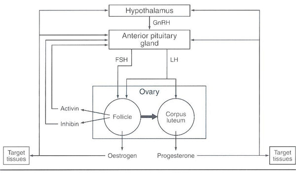
Figure 2. Hypothalamic-pituitary-ovary axis showing hormonal secretion pathways (from Gary C.W. England. in Canine and Feline Reproduction and Neonatology. BSAWA 2010.)
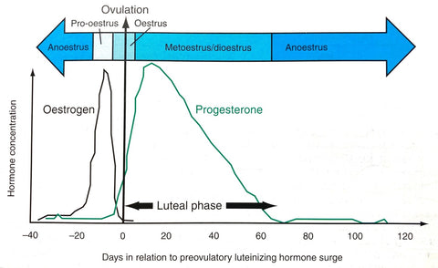
One specific feature of the canine estrus cycle is the so-called preovulatory luteinization. At the time of LH peak, the follicles start to secrete progesterone. After ovulation, the follicle turns into a corpus luteum and progesterone concentration keeps increasing. The rise continues up to 35 days and then slowly declines at the end of diestrus. The progesterone profile is very similar both in the pregnant and nonpregnant dog; one more specific feature in the canine estrus cycle. In anestrus, the concentrations of estrogen, LH and progesterone are low (Figure 3.).

Figure 3. Days in relation to preovulatory luteinizing hormone surge
Defining the time of the ovulation is essential for a successful breeding. After the onset of the vaginal bleeding, the ovulation occurs on average in 11 days. However, there is a large variation from 3 to 31 days. The behavioral signs of the estrus do not correlate well with the time of the ovulation. The bitch may show standing heat and be willing to mate even before the ovulation and may also allow mating even 10 days after the ovulation. Mating before the ovulation may result in pregnancy if the semen quality is good and the sperm cells stay viable until the egg cells have reached maturity after the ovulation. The egg cells stay fertile for about 5 days after the ovulation. If the mating happens after that, no pregnancy will result.
Swelling of the vulva and vaginal wall in the estrus is caused by estrogen. The degree of estrogen secretion can be estimated by assessing the changes in the vaginal epithelium. Estrogen influence can be seen as the keratinization of the epithelial cells which reaches its maximum of >80% keratinized superficial cells around the LH peak. However, the variation is wide and the optimal time of breeding cannot be based only on the vaginal cytology. (Figure 4.).

Figure 4. Keratinized vaginal epithelial cells in estrus (picture: Merja Dahlbom)
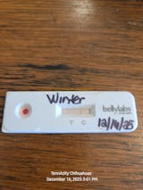
To get more precise diagnosis of the ovulation, the progesterone measurement is today the diagnostic tool of choice, preferably together with the clinical signs and vaginal cytology. Due to the preovulatory luteinization, progesterone value starts its rise two days before the ovulation. The progesterone value during the LH peak is around 2 ng/ml. At the time of the ovulation progesterone concentration has reached 5 ng /ml (Figure 5.).
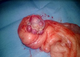
Figure 5. Corpora lutea in the dog ovary (Picture: Merja Dahlbom)
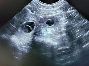
Ultrasonography offers an even more precise way for the ovulation diagnosis. In the study done in labrador retrievers, the day of ovulation was detected in 98.7% of follicles detected (24 cycles). Most of the ovulations (81.5%) occurred two to three days after the LH peak.
Combination of the clinical signs, vaginal cytology, progesterone measurement and ultrasonography gives a good estimate of the ovulation. Ultrasonography is especially useful when monitoring the estrus cycle in the infertile dogs.
References
Sebastian Arlt. Canine ovulation timing: A survey on methodology and an assessment on reliability of vaginal cytology. Reprod Domest Anim. 2018 Nov;53 Suppl 3:53-62.
Concannon, P.W.; Hansel, W.; Visek, W.J. The ovarian cycle of the bitch: plasma Estrogen, LH and progesterone. Biol. Reprod. 1975, 13,112-121.
Concannon, P.W.; McCann, J.P.; Temple, M. Biology and endocrinology of ovulation, pregnancy and parturition in the dog. J. Reprod. Fertil. Suppl. 1989, 39, 3-25.
Concannon, P.W. Reproductive cycles of the domestic bitch. Anim. Reprod. Sci. 2011, 124, 200-210.
England G., von Heimendahl A., BSAVA manual of Canine and Feline Reproduction and Neonatology, BSAVA, 2.ed. 2010.
T Hori1, T Tsutsui, Y Amano, P W Concannon.Ovulation day after onset of vulval bleeding in a beagle colony. Reprod Domest Anim , 2012 Dec;47 Suppl 6:47-51.
Kowalewski, M.P.; Ihle, S.; Siemieniuch, M.J.; Gram, A.; Boos, A.; Zduńczyk, S.; Fingerhut, J.; Hoffmann, B.; Schuler, G.; Jurczak, A.; Domosławska, A.; Janowski, T. Formation of the early canine CL and the role of prostaglandin E2 (PGE2) in regulation of its function: An in vivo approach. Theriogenology 2015, 83, 1038-1047.
Reynaud, K.; Saint-Dizier, M.; Tahir, M.Z.; Havard, T.; Harichaux, G.; Labas, V.; Thoumire, S.; Fontbonne, A.; Grimard, B.; Chastant-Maillard, S. Progesterone Plays a Critical Role in Canine Oocyte Maturation and Fertilization. Biol. Reprod. 2015, 93, 1-9.
K Reynaud, A Fontbonne, M Saint-Dizier, S Thoumire. C Marnier, M Z Tahir, T Meylheuc, S Chastant-Maillard, Folliculogenesis, ovulation and endocrine control of oocytes and embryos in the dog Reprod Domest Anim. 2012 Dec;47 Suppl 6:66-9.
Mei Tsuchida, Nako Komura, Tatsuya Yoshihara, Yuta Kawasaki, Daichi Sakurai, Hiroshi Suzuki. Ultrasonographic observation in combination with progesterone monitoring for detection of ovulation in Labrador Retrievers. Reprod Domest Anim . 2022 Feb;57(2):149-156.


